Induced Native Phage Therapy (INPT) and related Inducen® formulations are intended to function as an important key component of a broader, integrative, non-cytotoxic therapeutic strategy. They are not proposed as standalone cancer treatment, but as adjunctive interventions designed to address contributory microbial drivers of oncogenesis while preserving commensal microbiome integrity.
List of 10 Bacterial Infections That Can Instigate and Perpetuate Cancer
Below is an updated list of various bacterial infections known or suspected to instigate cancer, based on our previous discussions and peer-reviewed research from the web search results. The list focuses on key bacteria, the associated cancer types, and the mechanisms by which they initiate or promote carcinogenesis. These mechanisms often involve chronic inflammation, DNA damage, immune evasion, and oncogenic signaling pathways. Note that while some associations are causative (e.g., Helicobacter pylori as a Class I carcinogen by WHO¹), others are correlative and require further validation.
This list is not exhaustive but highlights some of the most well-established associations. Bacterial infections contribute to ~15–20% of global cancers³⁶,³⁷, primarily through chronic inflammation and DNA damage. For INPT targeting, focusing on these bacteria could disrupt their oncogenic roles across cancer types.
Implications of Induced Native Phage Therapy for Oncology: A Microbiome-Centered Hypothesis
An expanding body of evidence indicates that many solid tumors harbor distinct, metabolically active microbial communities within the tumor microenvironment (TME)³⁸,³⁹. These tumor-associated bacteria are increasingly recognized as contributors to oncogenesis and tumor persistence through mechanisms that include chronic inflammation, immune evasion, altered antigen presentation, and metabolic reprogramming⁴⁰. Notably, enrichment of specific bacterial taxa—such as Fusobacterium nucleatum—has been documented across multiple malignancies⁴¹–⁴³.
In colorectal cancer, F. nucleatum has been shown to promote tumor progression through adhesin-mediated interactions (e.g., FadA) that activate β-catenin signaling, as well as immune evasion via Fap2-mediated engagement of inhibitory receptors on natural killer and T cells²⁸–³³. Similar microbial signatures have been identified in other tumor types⁴¹.
These observations motivate investigation of therapeutic strategies that selectively disrupt tumor-associated bacteria without broadly damaging host tissues or the commensal microbiome. Bacteriophages are uniquely suited for this role, as they exhibit strain-restricted host specificity and can replicate at sites where susceptible bacteria are present⁴⁴–⁴⁷. Preclinical and translational studies have demonstrated that phage-mediated targeting of tumor-associated bacteria can alter immune infiltration, reduce pro-tumorigenic inflammation, and enhance responsiveness to conventional therapies in experimental systems⁴⁸.
Induced Native Phage Therapy (INPT) extends this conceptual framework by proposing a non-pharmacological method intended to recruit bacteriophages already present within the host phageome, rather than administering exogenous phage preparations. Within this model, strain-targeted electromagnetic signature sets are selected and encoded via Biospectral Emission Sequencing (BES) with the objective of selectively engaging microbial targets and facilitating conditions permissive for phage-mediated killing. Importantly, this framework does not require that all tumor-associated bacteria be eliminated, nor does it presume direct cytotoxicity toward malignant cells; rather, it hypothesizes that selective microbial reduction may indirectly remodel the TME by attenuating inflammatory signaling, disrupting biofilm-like microbial niches, and restoring immune surveillance as represented in Figure 1.
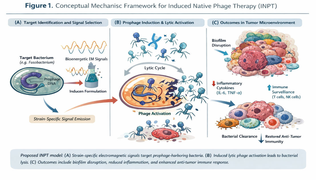
Figure 1 illustrates the proposed, hypothesis-consistent mechanism by which Induced Native Phage Therapy (INPT) may selectively reduce pathogenic or tumor-associated bacterial populations through recruitment of endogenous bacteriophages.
At present, the relevance of INPT to oncology remains a hypothesis grounded in biological plausibility rather than demonstrated clinical efficacy inside of IRB-supported research protocols. While infection-focused studies suggest rapid, strain-selective microbial clearance consistent with phage-mediated dynamics⁴⁴–⁴⁷, direct evidence of prophage induction, lytic transition, and phage amplification within tumor-associated bacteria has not yet been established, due to the nature of limited imaging capabilities of in vivo phage/microbe interactions. Accordingly, oncology applications of INPT is being evaluated through staged investigation, including tumor-resolved microbiome and phageome profiling, assessment of prophage induction markers, spatial immune-microbial mapping, and controlled clinical studies with predefined oncologic endpoints.
Nevertheless, the convergence of three independently established observations—(i) the detection of the presence of tumor-associated bacteria across multiple cancers³⁸–⁴³, (ii) the capacity of bacteriophages to be induced through INPT to selectively eliminate specifically targeted bacterial populations and amplify in situ⁴⁴–⁴⁷, and (iii) the sensitivity of tumor immune dynamics to microbial cues⁴⁰,⁴⁹—supports the exploration of INPT as a microbiome-modulating adjunct rather than a stand-alone anticancer therapy. If validated, such an approach could complement existing modalities by addressing microbial drivers of immune suppression and therapeutic resistance that are not directly targeted by the conventional cytotoxic or immunologic agents of oncology.
References
- International Agency for Research on Cancer. Schistosomes, Liver Flukes and Helicobacter pylori. IARC Monographs on the Evaluation of Carcinogenic Risks to Humans, Volume 61 (1994).
- Polk, D.B. & Peek, R.M. Helicobacter pylori: gastric cancer and beyond. Nature Reviews Cancer 10, 403–414 (2010).
- Hatakeyama, M. Helicobacter pylori CagA and gastric cancer: a paradigm for hit-and-run carcinogenesis. Cell Host & Microbe 15, 306–316 (2014).
- Ohnishi, N. et al. Transgenic expression of Helicobacter pylori CagA induces gastric carcinoma in mice. Proceedings of the National Academy of Sciences of the United States of America 105, 1003–1008 (2008).
- Nath, G. et al. Chronic typhoid carriage and carcinoma of the gallbladder. European Journal of Cancer Prevention 6, 557–559 (1997).
- Scanu, T. et al. Salmonella manipulation of host signaling pathways provokes cellular transformation associated with gallbladder carcinoma. Cell Host & Microbe 17, 763–774 (2015).
- Boleij, A. et al. Clinical importance of Streptococcus gallolyticus infection among colorectal cancer patients. Clinical Infectious Diseases 53, 870–878 (2011).
- Abdulamir, A.S., Hafidh, R.R. & Abu Bakar, F. The association of Streptococcus bovis/gallolyticus with colorectal tumors. Journal of Experimental & Clinical Cancer Research 30, 11 (2011).
- Kumar, R. et al. Streptococcus gallolyticus promotes colorectal tumor development. PLoS Pathogens 13, e1006440 (2017).
- Tjalsma, H. et al. A bacterial driver–passenger model for colorectal cancer. Microbial Ecology in Health and Disease 23, 18588 (2012).
- Pasquereau-Kotula, E. et al. Virulence of Streptococcus gallolyticus in colorectal cancer. Future Microbiology 13, 1179–1193 (2018).
- Littman, A.J. et al. Chlamydia pneumoniae infection and risk of lung cancer. Cancer Epidemiology, Biomarkers & Prevention 13, 1624–1630 (2004).
- Zhan, P. et al. Chlamydia pneumoniae infection and lung cancer risk: a meta-analysis. Tumor Biology 32, 887–896 (2011).
- Nougayrède, J.P. et al. Escherichia coli induces DNA double-strand breaks in eukaryotic cells. Science 313, 848–851 (2006).
- Arthur, J.C. et al. Intestinal inflammation targets cancer-inducing activity of the microbiota. Science 338, 120–123 (2012).
- Cuevas-Ramos, G. et al. Escherichia coli induces DNA damage in vivo and triggers genomic instability. Proceedings of the National Academy of Sciences USA 107, 11537–11542 (2010).
- Pleguezuelos-Manzano, C. et al. Mutational signature in colorectal cancer caused by genotoxic pks+ Escherichia coli. Nature 580, 269–273 (2020).
- Dejea, C.M. et al. Patients with familial adenomatous polyposis harbor colonic biofilms containing tumorigenic bacteria. Science 359, 592–597 (2018).
- Wu, S. et al. A human colonic commensal promotes colon tumorigenesis via activation of T helper type 17 T cell responses. Nature Medicine 15, 1016–1022 (2009).
- Chung, L. et al. Bacteroides fragilis toxin coordinates a pro-carcinogenic inflammatory cascade. Cell Host & Microbe 23, 203–214 (2018).
- Sears, C.L. Enterotoxigenic Bacteroides fragilis: a rogue among symbiotes. Clinical Microbiology Reviews 22, 349–369 (2009).
- Housseau, F. et al. Microbiota and colon cancer: Bacteroides fragilis enters the fray. Immunity 39, 603–605 (2013).
- Gallimidi, A.B. et al. Periodontal pathogens promote tumor progression in an oral-specific chemical carcinogenesis model. Proceedings of the National Academy of Sciences USA 112, E2488–E2497 (2015).
- Michaud, D.S. et al. Periodontal disease, tooth loss, and cancer risk. The Lancet Oncology 9, 550–558 (2008).
- Whitmore, S.E. & Lamont, R.J. Oral bacteria and cancer. PLoS Pathogens 10, e1003933 (2014).
- Smith, J.S. et al. Chlamydia trachomatis and invasive cervical cancer. Journal of the National Cancer Institute 96, 482–489 (2004).
- Madeleine, M.M. et al. Co-infection with Chlamydia trachomatis and HPV increases cervical cancer risk. Cancer Epidemiology, Biomarkers & Prevention 16, 128–135 (2007).
- Rubinstein, M.R. et al. Fusobacterium nucleatum promotes colorectal carcinogenesis by modulating E-cadherin/β-catenin signaling. Cell Host & Microbe 14, 195–206 (2013).
- Kostic, A.D. et al. Fusobacterium nucleatum potentiates intestinal tumorigenesis. Cell Host & Microbe 14, 207–215 (2013).
- Gur, C. et al. Binding of the Fap2 protein to human inhibitory receptor TIGIT protects tumors. Immunity 42, 344–355 (2015).
- Bullman, S. et al. Analysis of Fusobacterium persistence and antibiotic response in colorectal cancer. Science 358, 1443–1448 (2017).
- Mitsuhashi, K. et al. Association of Fusobacterium species in pancreatic cancer tissues. Clinical Cancer Research 21, 5467–5473 (2015).
- Yamamura, K. et al. Fusobacterium nucleatum in gastroenterological cancers. Cancer Science 108, 100–105 (2017).
- Alpern, R.J. & Dowell, V.R. Clostridium septicum infections and malignancy. American Journal of Medicine 46, 70–77 (1969).
- Kornbluth, A.A. et al. Clostridium septicum infection and colorectal carcinoma. Annals of Internal Medicine 111, 547–550 (1989).
- de Martel, C. et al. Global burden of cancers attributable to infections in 2008. The Lancet Oncology 13, 607–615 (2012).
- Plummer, M. et al. Global burden of cancers attributable to infections in 2012. The Lancet Global Health 4, e609–e616 (2016).
- Nejman, D. et al. The human tumor microbiome is composed of tumor type–specific intracellular bacteria. Science 368, 973–980 (2020).
- Poore, G.D. et al. Microbiome analyses of blood and tissues suggest cancer diagnostic potential. Nature 579, 567–574 (2020).
- Zitvogel, L. et al. The microbiome in cancer immunotherapy. Science 359, 1366–1370 (2018).
- Castellarin, M. et al. Fusobacterium nucleatum infection is prevalent in colorectal carcinoma. Genome Research 22, 299–306 (2012).
- Mima, K. et al. Fusobacterium nucleatum and T cell infiltration in colorectal carcinoma. Journal of the American Medical Association Oncology 1, 653–661 (2015).
- Brennan, C.A. & Garrett, W.S. Fusobacterium nucleatum — symbiont, opportunist and oncobacterium. Nature Reviews Microbiology 17, 156–166 (2019).
- Abedon, S.T. et al. Phage therapy pharmacology. Clinical Microbiology Reviews 24, 318–348 (2011).
- Dedrick, R.M. et al. Engineered bacteriophages for treatment of drug-resistant infection. Nature Medicine 25, 730–733 (2019).
- Górski, A. et al. Phages and immunomodulation. Frontiers in Immunology 8, 981 (2017).
- Little, J.W. Lysogeny and prophage induction. Annual Review of Microbiology 59, 73–93 (2005).
- Hodyra-Stefaniak, K. et al. Mammalian immune responses to bacteriophages. Frontiers in Immunology 6, 221 (2015).
- Gopalakrishnan, V. et al. Gut microbiome modulates response to anti–PD-1 immunotherapy in melanoma patients. Science 359, 97–103 (2018).